
Nuclear Medicine and Its Applications in Gastroenterology
Medical imaging plays a large role in the way physicians study the body and diagnose conditions. Nuclear medicine offers a distinct approach, and it focuses on how organs function rather than only how they look. For people who experience digestive symptoms, this branch of imaging can help doctors gather detailed information about the stomach, intestines, liver, and related organs.
What Is Nuclear Medicine?
Nuclear medicine is a branch of medical imaging that uses small amounts of radioactive material to examine how the body works. These materials, often called radiotracers, are given to the patient before imaging begins. Once inside the body, a radiotracer travels to a specific organ or tissue and releases a low level of energy. A special camera then detects that energy, and it produces images that show activity inside the body.
The main difference between nuclear medicine and more familiar tests, such as standard X-rays, is the type of information provided. An X-ray shows the shape and structure of an area. Nuclear medicine focuses on function, meaning it reveals how well an organ is performing its normal tasks. This functional view can help physicians identify changes that structural images may not display. The amount of radioactive material used in these studies is typically low, and the tracers are designed to leave the body over time.
What Is It Used For?
Gastroenterology deals with the digestive system, which includes the esophagus, stomach, intestines, liver, gallbladder, and pancreas. Nuclear medicine supports this field by helping physicians study how these organs move and process substances. One common example is the gastric emptying study. This test measures how quickly food leaves the stomach and moves into the small intestine. A patient eats a meal that contains a small amount of radiotracer, and the camera records the movement over time. The results can help a physician understand digestive patterns. Another application involves the liver and gallbladder. A hepatobiliary study examines how bile flows through these organs. This type of imaging can support the evaluation of gallbladder function and the pathways that carry bile.
What Should You Expect?
A nuclear medicine appointment usually follows a straightforward sequence. Before the visit, your care team may give you specific instructions, such as avoiding food for a set period or pausing certain medications. The radiotracer is then administered, and it is most often done through an injection or swallowed orally. Imaging begins shortly after the tracer is given in some cases. In others, you may wait a period of time so the tracer can reach the target organ.
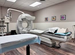
During imaging, you will lie on a table while the camera moves near your body to capture pictures. The process is generally painless. You will be asked to remain still so the images stay clear. Depending on the study, the imaging portion can last from several minutes to a few hours, and some tests require more than one set of pictures across a longer window.
After the appointment, most people return to their usual activities. The radiotracer fades from the body naturally, and staff may suggest drinking water to support this process. A specialist can review the images and share the findings with the physician who ordered the study.
Seek Professional Radiology Services
Nuclear medicine gives physicians a valuable tool for studying the function of the digestive system. These studies focus on how organs perform their tasks, and they may complement other tests and support a better understanding of a person’s health. If a digestive concern calls for functional imaging, trained radiology professionals can perform the study and work alongside your gastroenterologist.